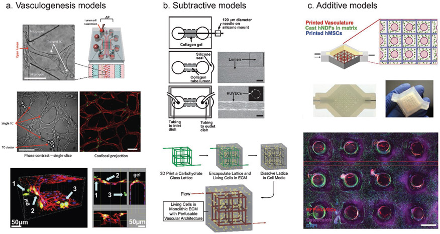
Figure 2.
Common methods to vascularize biomaterials. a) Vasculogenesis models induce sprouting of ECs into a biomaterial. A schematic of a commonly used vasculogenesis model is displayed in the top right. This model allows visualization of trapped tumor cells (top left and middle, with arrows indicating trapped tumor cells or clusters of tumor cells), as well as extravasated tumor cells (bottom image arrows). b) Subtractive models create an empty space in a material that can be lined with ECs. In the top image, a collagen gel was polymerized over a needle, which was then removed, and the remaining empty space was perfused with ECs to create a single channel. In the bottom image, a carbohydrate lattice was 3D printed and then encapsulated in ECM mimic. The lattice can then be dissolved, with the resulting empty space perfused and lined with ECs. c) Additive models (3D bioprinting) directly deposit the biomaterial of choice as well as multiple cell types to build a model tissue from the ground up. Here, a bioprinter was used to print ECs, fibroblasts, and stem cells to create a thick vascularized tissue. (a) Bottom: reproduced with permission.[43] Copyright 2013, PLoS One. Top and middle: reproduced with permission.[85] Copyright 2017, Springer Nature. (b) Top: reproduced with permission.[36] Copyright 2006, Elsevier. Bottom: reproduced with permission.[35] Copyright 2012, Springer Nature. (c) Reproduced with permission.[54] Copyright 2016, National Academy of Sciences.

