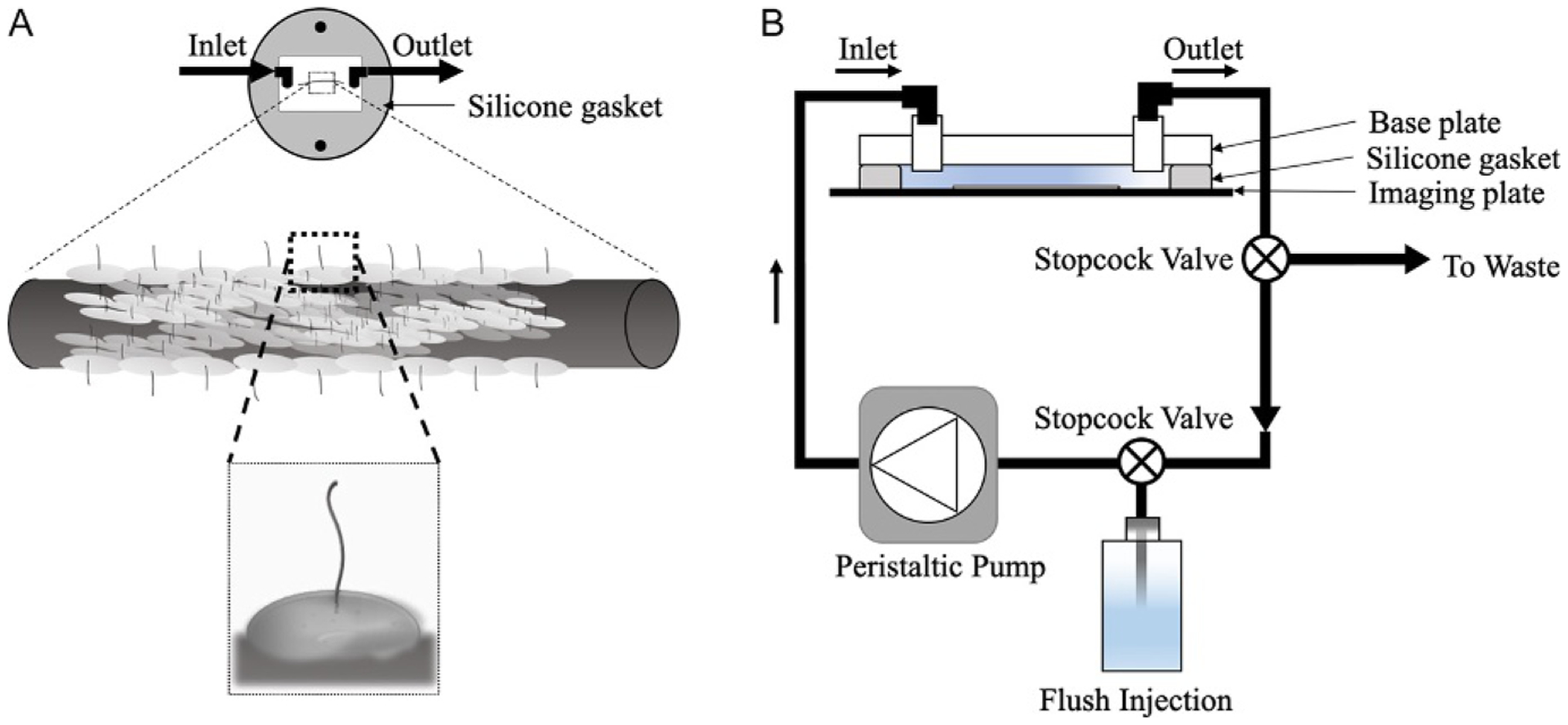
FIG. 3.

Microwire with cells in a perfusion chamber. (A) In the diagram, the gasket is placed on the imaging dish in a manner to orient the microwire parallel to laminar flow. Using lower magnifications, we can observe cells growing on the wire edge. At a higher magnification, the cilium protruding outward should become even clearer. We will focus on a single cell and capture Ca2+ data in response to fluid flow. (B) Schematic of the experimental setup with stopcock valves to load and unload buffers into circulation.
