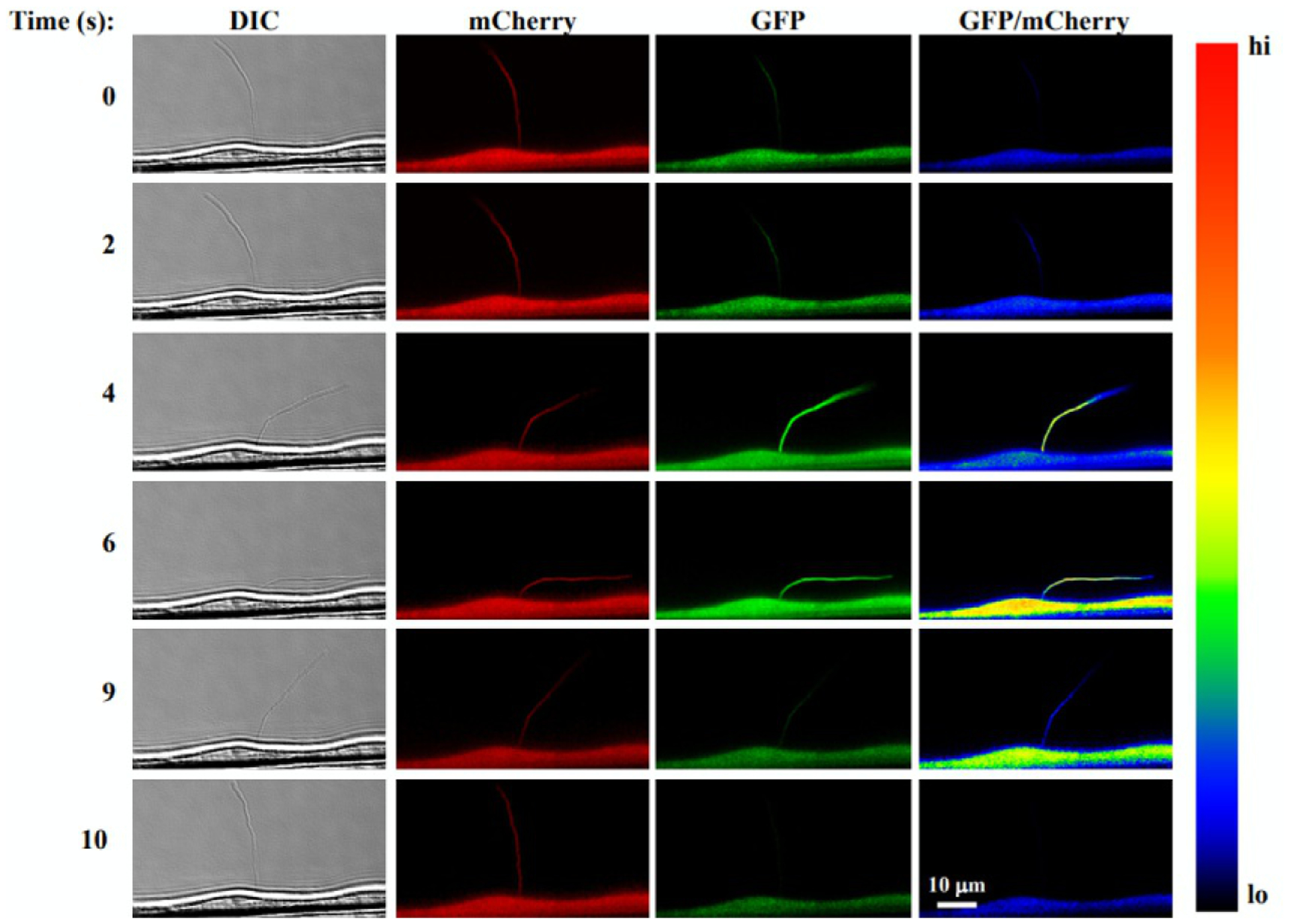
FIG. 4.

Single-live cell and cilia imaging with 5HT6-mCherry-G-GECO1.0. The images show (from left to right column) DIC used for tracking a cilium, mCherry fluorescent ciliary marker, the Ca2+ sensitive GECO1.0 and an EGFP/mCherry ratio pseudocolored to show Ca2+ levels. When fluid flow is applied (time series from top to bottom), the cilium bends inducing a Ca2+ increase in both the cytoplasm and cilioplasm.
Adapted with permission from Pala, R., Mohieldin, A. M., Shamloo, K., Sherpa, R. T., Kathem, S. H., Zhou, J., et al. Personalized nanotherapy by specifically targeting cell organelles to improve vascular hypertension. Nano Letters, 19, 904–914. Copyright 2018 American Chemical Society.
