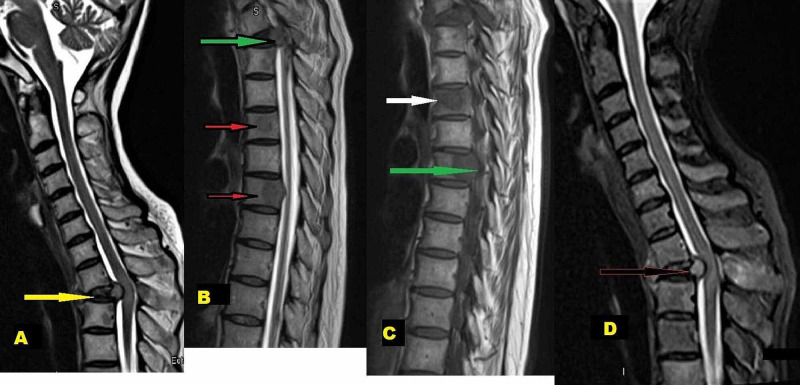
Figure 3. MRI Spine.
A) (Para-sagittal view) T2-weighted images showing compression at T1-T2 spinal level (yellow arrow)
B) Multiple spinal bony metastasis (red arrows) with compression on the anterior spinal cord (green arrow)
C) T1-weighted MRI images showing body metastatic (white arrow) and pedicles metastatic involvement (green arrow)
D) Extradural compression on the cord pushed posteriorly (arrow)

