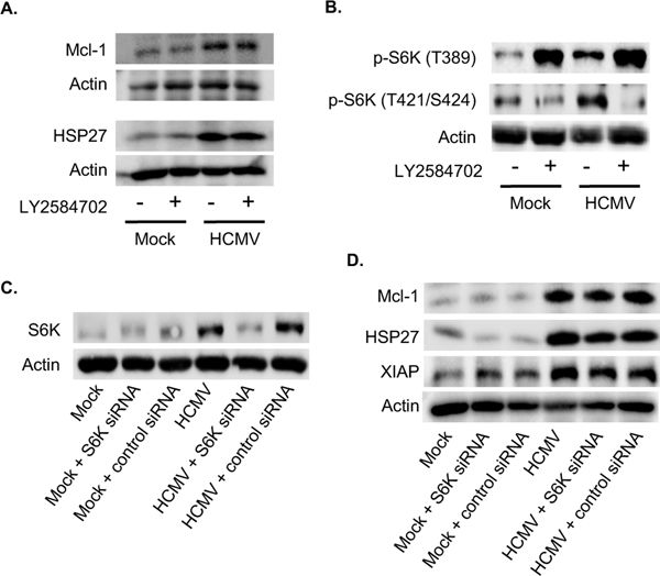
Fig. 3. Activation of S6K is not responsible for the increased expression of Mcl-1 and HSP27 following HCMV infection.
(A-B) Monocytes were pretreated with LY2584702 (a S6K inhibitor) for 1 h, then mock or HCMV infected for 24 h. (A) Mcl-1 and HSP27 or (B) pS6K (T389) and pS6K (T421/S424) were detected by immunoblotting. (C-D) Monocytes were transfected with a scrambled control siRNA or S6K siRNA and incubated for 24 h. Following incubation, cells were mock infected or HCMV infected for an additional 24 h. (C) S6K or (D) Mcl-1, HSP27 and XIAP expression were determined by immunoblotting. (A-D) Membranes were reprobed for β-actin as a loading control. Data are representative of 3–6 independent blood donors.

