FIGURE 3.
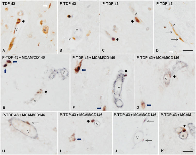
TDP-43 immunoreactivity in frontal cortex area 8 and subcortical white matter in FTLD-TDP type A. TDP-43 immunoreactivity is observed as small round inclusions (A, C) or as elongated or punctate inclusions (B, D) associated with the wall of blood vessels. Double-labeling immunohistochemistry with P-TDP-43 Ser403-404 (brown) and MCAM/CD146 (dark blue) antibodies shows the relationship of these inclusions with cells of the blood vessel walls (E–K). Some of them were located within the blood vessel walls (E, G, K), but others were bounded to the external surface of the blood vessels (F, H, I, J). Short thick arrows indicate round inclusions, and thin arrows elongated or punctate inclusions in blood vessels; long thick arrows, neuronal TDP-43-immunoreactive cytoplasmic inclusions in neurons; white arrow, TDP-43 containing neurites; asterisk, preserved nuclear TDP-43 immunoreactivity in one neuron; V: blood vessel. Paraffin sections, lightly counterstained with hematoxylin, scale bars: A–D = 30 µm; E–K = 25 µm.

