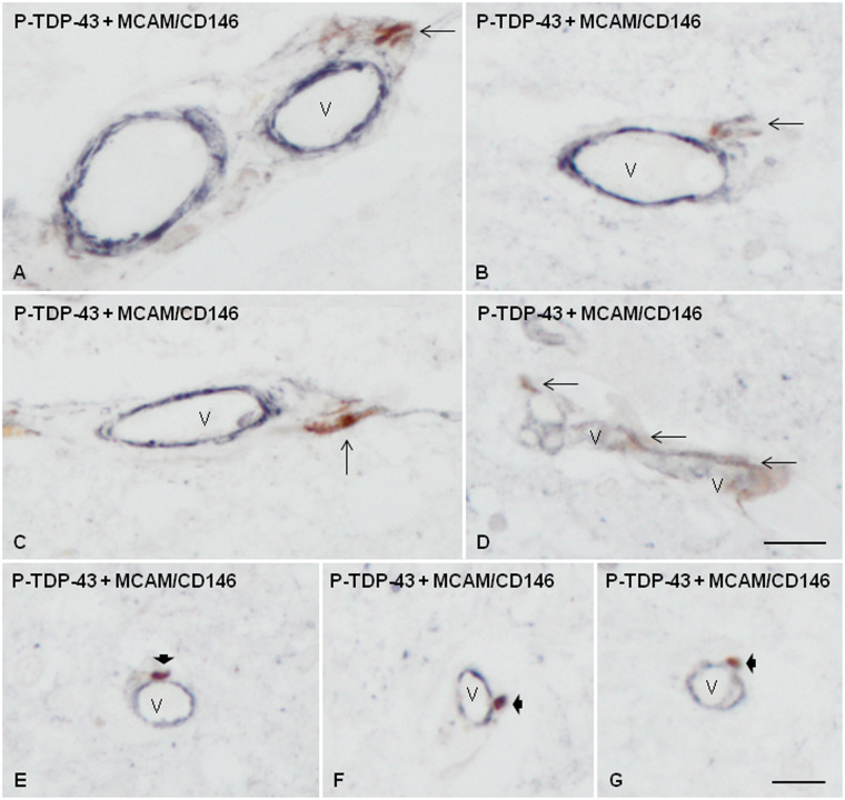
FIGURE 4.
Double-labeling immunohistochemistry with antibodies P-TDP-43 Ser403-404 (brown) and MCAM/CD146 (dark blue) showing P-TDP-43-immunoreactive elongated (thin arrows) or globular (short thick arrows) inclusions in association with the blood vessels (V) in the spinal cord of sALS cases. Some elongated large inclusions were localized in the external layer or the perivascular space (A–C); others in the blood vessel wall (D), and others were attached to the external surface of small blood vessels (E–G). Paraffin sections, scale bars = 20 µm, excepting D, scale bar = 25 µm.

