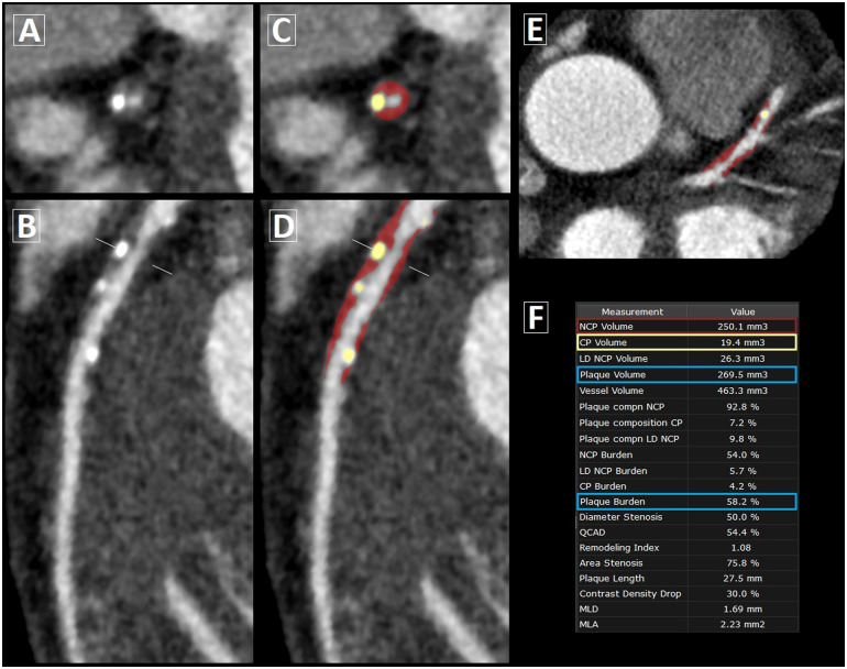
Figure 2.
Semi-automated coronary plaque analysis on CCTA. Case example of plaque quantification in a 67-year-old male patient with stable CAD. (A) Cross-section and (B) curved multi-planar view of mixed plaque in the proximal-to-mid left anterior descending artery. (C and D) Corresponding views and (E) axial slice following semi-automated plaque analysis, with non-calcified plaque (NCP) in red overlay and calcified plaque (CP) in yellow overlay. (F) Plaque volume = NCP volume + CP volume; plaque burden = plaque volume × 100%/analysed vessel volume.

