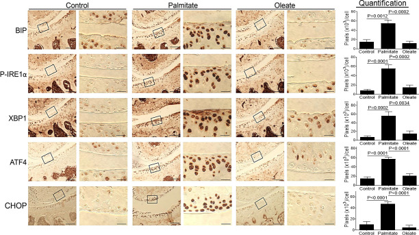
Fig 2. Dietary palmitate induces ER stress in mouse knee cartilage.
Mice were fed with a control, palmitate, or oleate diet (n = 15 per diet group) for 20 weeks. Joints were collected, processed and sectioned (one section knee/mouse) for immunohistochemical staining. Coronal sections of mouse knee joints were analyzed for ER stress markers including BIP, P-IRE1α, XBP1, ATF4, and CHOP. Images on left panels in each pair of columns are of low magnification (Scale bars: 100 μm), and the tibia is in the lower half of images. The areas inside the small rectangles were magnified and displayed in right panels (scale bars: 20 μm). All immunohistochemical data were quantified with correction for cell numbers and statistically analyzed. The bars were obtained from the analysis of 3 mice per group (n = 3), and the data are expressed as the average pixel numbers (×103) per cell ± standard deviation.

