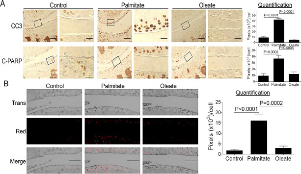
Fig 3. Dietary palmitate induces chondrocyte apoptosis in mouse knee cartilage.
Mice were fed a control, palmitate, or oleate diet (n = 15 per diet group) for 20 weeks. Joints were collected, processed, and sectioned. We used a single section from the right knee/mouse (n = 3) for immunohistochemical and TUNEL staining. (A) Coronal sections of mouse knee joints were analyzed immunohistochemically for apoptosis markers CC3 and C-PARP. Images on left panels in each pair of columns are of low magnification (Scale bars: 100 μm), and the tibia is in the lower half of images. The areas inside the small rectangles were magnified and displayed in right panels (scale bars: 20 μm). (B) Coronal sections of mouse knee joints were evaluated by TUNEL staining. Scale bar, 100 μm. All immunohistochemical and TUNEL data were quantified with correction for cell numbers and statistically analyzed. The bars were obtained from the analysis of 3 mice per group (n = 3), and the data are expressed as the average pixel numbers (×103) per cell ± standard deviation.

