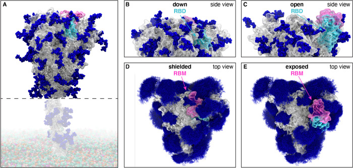
Figure 1.
Glycosylated spike RBD “down” and “open” conformations. (A) The SARS-CoV-2 spike head (gray) with glycans (dark blue) as simulated, with the stalk domain and membrane (not simulated here, but shown in transparent for completeness). RBD shown in cyan, receptor binding motif (RBM) in pink. Side view of the “down” (shielded, B) and “open” (exposed, C) RBD. Top view of the closed (shielded, D) and “open” (exposed, E) RBM. Composite image of glycans (dark blue lines) shows many overlapping snapshots of the glycans over the microsecond simulations.

