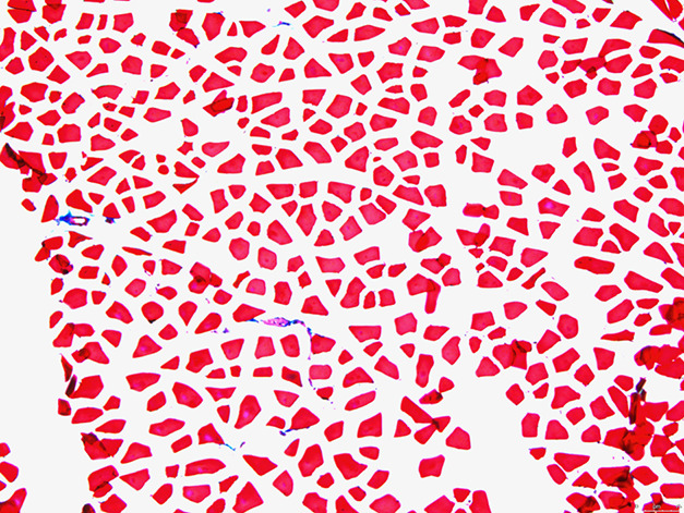
Fig. 3.

This histologic image (at 10 × magnification) is of the quadriceps muscle of a mouse 9 months after irradiation, treated with the TGF-ß inhibitor (1 mg/kg daily for 8 weeks) after radiation. Fresh-frozen tissue was stained with Masson’s trichrome (red indicates muscle; blue indicates fibrosis).
