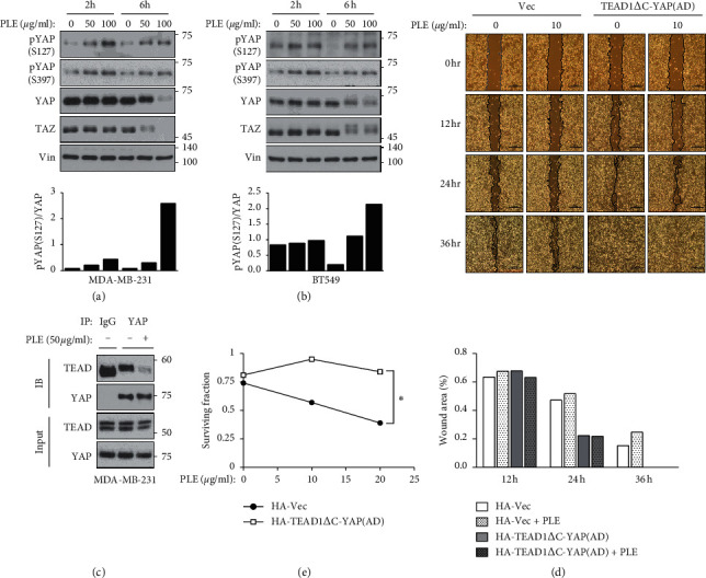
Figure 5.

Constitutively active YAP-TEAD rescued PLE-induced inhibition of cancer cell migration and proliferation. (a, b) To determine the effect of PLE on the YAP phosphorylation, MDA-MB-231 and BT549 cells were treated with PLE (25 and 50 μg/mL) for 6 and 12 h. Cell lysates were subjected to immunoblotting with the indicated antibodies. The bar graphs show the pYAP (S127) amounts normalized to total YAP amounts for individual samples. (c) HEK293A cells were treated with PLE (50 μg/mL) for 6 h. Endogenous YAP/TAZ was immunoprecipitated, and the co-precipitated pan-TEAD was detected by western blot. (d) Mobility of MDA-MB-231 cells expressing the HA-vector or TEAD1ΔC-YAP (AD) was evaluated using the wound healing assay. The cells were wounded and treated with PLE (10 μg/mL) for 36 h in a serum-containing medium. At different time points (0, 12, 24, and 36 h), phase-contrast photographs of the wounds were taken and analyzed using ImageJ program. (e) MDA-MB-231 cells expressed with HA-vector or TEAD1ΔC-YAP (AD) were treated with PLE at concentrations of 10 and 25 μg/mL. The cells were applied to clonogenic growth assay after 10 days of the treatment. In the clonogenic growth assay, DMSO-treated cells served as a control. Each value is expressed as mean ± SEM from n = 3 per group. ∗p < 0.05, ∗∗p < 0.01; Student's t-test (unpaired, one-tailed) was used for statistical analysis. In Figure 5(b), the numbers of size maker depart from indicated lines.
