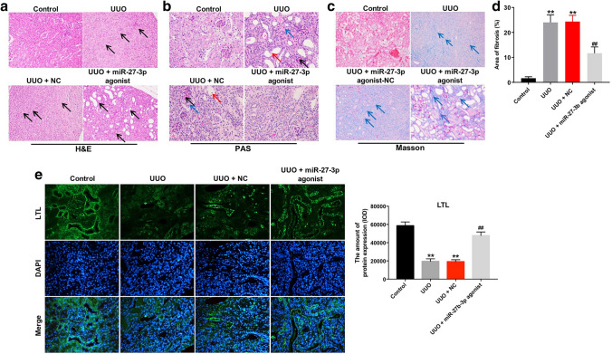
Fig. 4.
Overexpression of miR-27b-3p alleviated renal fibrosis in vivo. a Analysis of kidney injury in UUO kidneys by H&E staining (magnification, × 200). black arrows pointed to fibroblasts. b Representative photomicrographs of PAS-stained kidney sections (magnification, × 400). Black arrows pointed to glomerular mesangial matrix; red arrows pointed to exfoliation of renal tubular epithelial cells; blue arrows pointed to renal interstitial edema. c Analysis of collagen deposition and renal fibrosis by Masson’s trichrome staining (magnification, × 200). Blue arrow pointed to collagen fibers. d Total lung fibrotic area was measured by Image-Pro Plus. e Integrity of the glomerulotubular junction and proximal tubular mass were determined in LTL staining (magnification, × 200). **p < 0.01, compared with the control group. ##p < 0.01, compared with the UUO group

