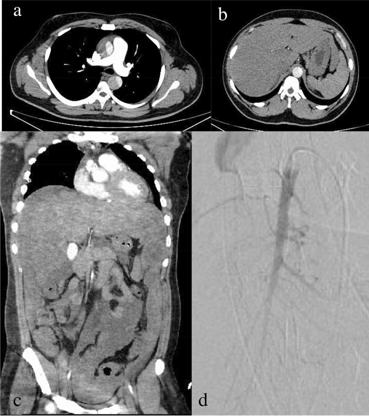
Fig. 1.
a, b Contrast-enhanced computed tomography (CT) showing type A aortic dissection and splenomegaly. c CT after the first operation showing ischemic small intestine showing a reduced contrast effect in peripheral region of superior mesenteric artery (SMA) and maintained contrast effect in the main trunk of SMA, along with the spleen and the liver infarct. d Angiography image showing reduced vessel filling in the peripheral region of the superior mesenteric artery

