Figure 2.
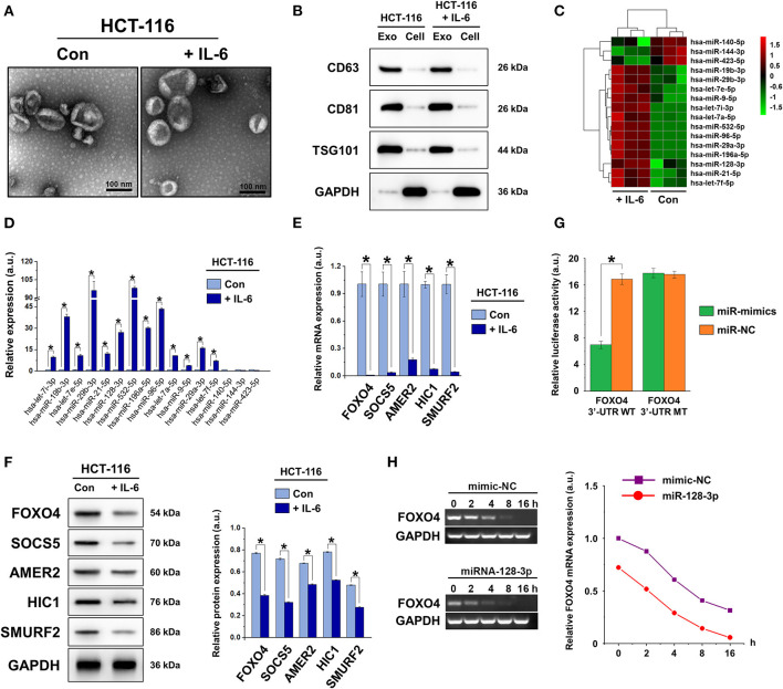
Characterization of exosomes derived from CRC cells. (A) TEM images showing small vesicles of ~40–100 nm in diameter, scale bar = 100 nm. (B) Western blot characterization of exosomal markers CD63, CD81, and TSG101 in isolated exosomes and cell lysates after extraction. (C) Heat map showing the changes in miRNA expression in exosomes isolated from non-induced (Con) and IL-6-induced HCT-116 cells. (D) qRT-PCR of exosomal miRNA expression in non-induced (Con) and IL-6-induced HCT-116 cells. (E) qRT-PCR and (F) western blot of the expression of miR-128-3p target genes FOXO4, SOCS5, AMER2, HIC1, and SMURF2 in non-induced (Con) and IL-6-induced HCT-116 cells. Protein expression was normalized to that of GAPDH. (G) Relative luciferase activity of reporter plasmids carrying WT or MUT FOXO4 3′-UTR in HCT-116 cells co-transfected with miR-128-3p mimics or its negative control (miR-NC). (H) Decay curve representative of FOXO4 mRNA degradation in HCT-116 cells carrying miR-128-3p mimics or mimic-NC. Data are shown as the mean ± SD (n = 3); *P < 0.05.

