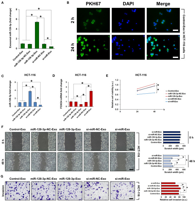
Figure 6.
Co-culture of HCT-116 cells with exosomes isolated from HCT-116 cells. HCT-116 cells were co-cultured with exosomes that were isolated from HCT-116 cells transfected with overexpression or interference vectors (or corresponding NC) of miR-128-3p. (A) qRT-PCR of relative expression of miR-128-3p in exosomes isolated from HCT-116 cells. (B) Indication of exosome uptake after 2 and 24 h in HCT-116 cells by fluorescence staining of PKH67. Green fluorescence represents PKH-labeled exosomes and blue fluorescence represent the cell nuclei. Scale bar = 50 μm. qRT-PCR of (C) miR-128-3p and (D) FOXO4 mRNA expression in HCT-116 cells co-cultured with exosomes. (E) Viability of HCT-116 cells was detected by CCK-8 assay after co-culture with exosomes. (F) Wound healing assay, scale bar = 200 μm. (G) Transwell invasion assay, scale bar = 100 μm. Data are shown as the mean ± SD (n = 3); *P < 0.05.

