FIGURE 2.

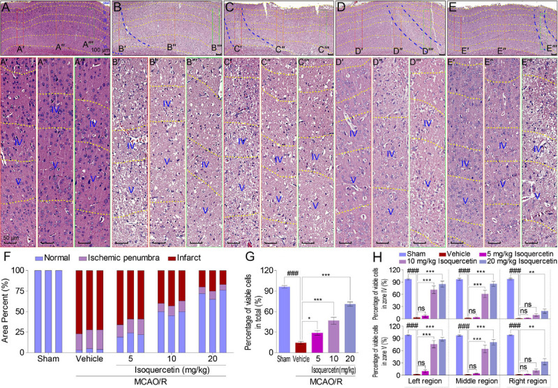
Effects of isoquercetin on cells in the parietal cortex of MCAO/R-induced rats by H&E staining. (A–E) Representative micrographs of the parietal cortex region in sham-operated, vehicle-treated, or isoquercetin-treated rats (A, sham; B, vehicle; C, 5 mg/kg isoquercetin; D, 10 mg/kg isoquercetin; E, 20 mg/kg isoquercetin). The parietal cortex was divided into seven layers (layer pial and I–VI). The penumbra region in the parietal cortex was marked with two blue dash lines. The left, middle, and right regions were marked with red-, orange-, and green-colored rectangles, and numbered as (A′–E″′). (A′–E″′) The zoom-in micrographs of marked regions in (A–E). The layers IV and V were signed for further quantitative analyses. (F) Quantitative analyses of the area percentage of ischemic penumbra area and infarct area in the parietal cortex of sham-operated, vehicle-treated, and isoquercetin-treated rats (n = 3). (G) Quantitative analyses of the percentage of viable cells in the parietal cortex of sham-operated, vehicle-treated, and isoquercetin-treated rats (n = 9). (H) Quantitative analyses of the percentage of viable cells within layers IV and V of the parietal cortex in sham-operated, vehicle-treated, and isoquercetin-treated rats (n = 3). Data were represented as mean ± SEM. ###p < 0.001 vs. sham group; *p < 0.05, **p < 0.01, and ***p < 0.001 vs. vehicle-treated group.
