
FIGURE 4.
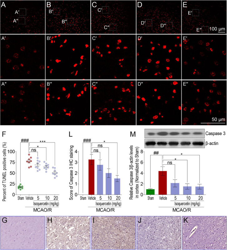
Effects of isoquercetin on cell apoptosis in the parietal cortex of sham-operated, vehicle-treated, and isoquercetin-treated rats. (A–E) Representative micrographs of cell apoptosis analyses of the parietal cortex by TUNEL staining (A, sham; B, vehicle; C, 5 mg/kg isoquercetin; D, 10 mg/kg isoquercetin; E, 20 mg/kg isoquercetin). (A′–E″) The zoom-in micrographs of marked regions in (A–E). (F) Quantitative analyses of apoptotic cells (featured by bright red signals) in cell population (n = 8). (G–K) Representative micrographs of IHC staining of Caspase 3 in the parietal cortex of sham-operated, vehicle-treated, and isoquercetin-treated rats (G, sham; H, vehicle; I, 5 mg/kg isoquercetin; J, 10 mg/kg isoquercetin; K, 20 mg/kg isoquercetin). (L) Assessment analyses of IHC score of Caspase 3 in the parietal cortex of sham-operated, vehicle-treated, and isoquercetin-treated rats (n = 4). (M) The protein level of Caspase 3 in the parietal cortex of rats were assayed by western blot analysis after MCAO/R injury and isoquercetin treatment. Data were expressed as mean ± SEM (n = 3). ##p < 0.01 and ###p < 0.001 vs. sham group; *p < 0.05 and ***p < 0.001 vs. vehicle-treated group.