
FIGURE 7.
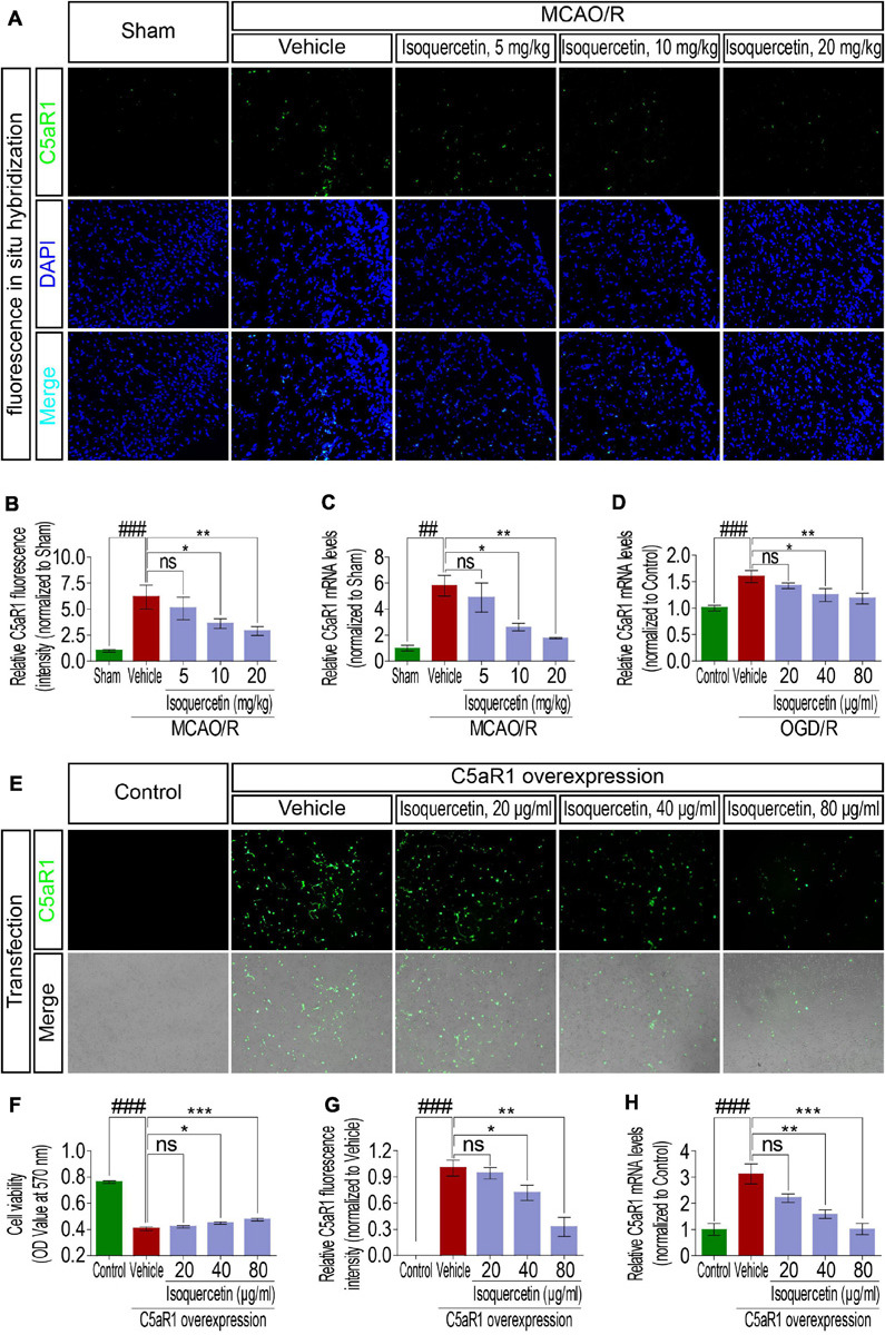
Effects of isoquercetin on C5aR1 expression in the parietal cortex of rats after MCAO/R and primary culture of cortical neurons after OGD/R injury and C5aR1 overexpression. (A) Representative micrographs of FISH of C5aR1 mRNA expression in the parietal cortex of sham-operated, vehicle-treated, and isoquercetin-treated rats. (B) The fluorescent intensity statistics of C5aR1 mRNA in the parietal cortex of sham-operated, vehicle-treated, and isoquercetin-treated rats (n = 4). (C) qPCR assay of C5aR1 mRNA expression levels in the parietal cortex of sham-operated, vehicle-treated, and isoquercetin-treated rats (n = 3). (D) qPCR assay of C5aR1 mRNA expression levels in control-, vehicle-, and isoquercetin-treated primary culture of cortical neurons (n = 3). (E) Representative micrographs of C5aR1 expression in primary culture of cortical neurons after over-expression and isoquercetin treatments. (F) Cell viability of cortical neurons after over-expression and isoquercetin treatments (n = 8). (G) The relative fluorescent intensity statistics of C5aR1 mRNA in cortical neurons after over-expression and isoquercetin treatments (n = 3). (H) qPCR assay of C5aR1 mRNA expression levels of cortical neurons after over-expression and isoquercetin treatments (n = 3). Results were expressed as mean ± SEM. ##p < 0.01 and ###p < 0.001 vs. sham or control group; *p < 0.05, **p < 0.01, and ***p < 0.001 vs. vehicle-treated group.