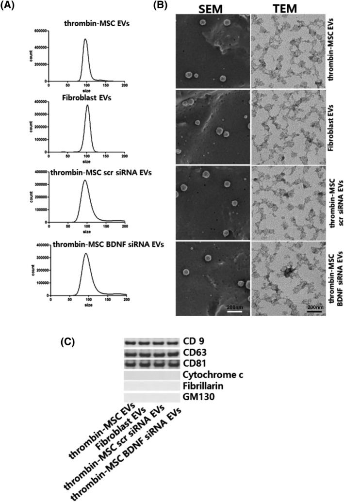
FIGURE 1.

Confirmation of extracellular vesicles (EVs). EVs were isolated from the cell culture media using ultracentrifugation. A, The size and number of EVs as measured using NanoSightNS300 and Nanoparticle Tracking Analysis software. B, Scanning electron micrograph (SEM) of EVs loaded on a polycarbonate membrane. Transmission electron micrograph (TEM) of EVs. EVs on copper grids and stained with uranyl acetate. Representative SEM (left) and TEM (right). C, Representative immunoblot for the organelle marker proteins in mesenchymal stem cell‐derived EVs, CD9, CD63, CD81, cytochrome C for mitochondria, fibrillarin for the nucleus, and GM130 for the Golgi
