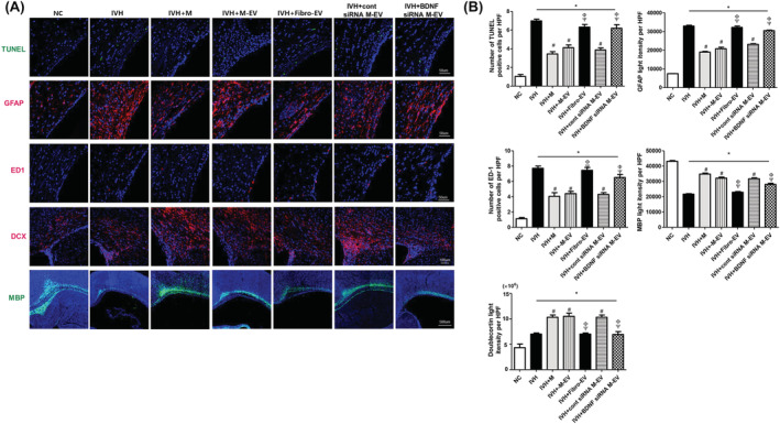
FIGURE 4.

BDNF knockdown in the extracellular vesicles abolished the therapeutic effects of MSCs in improving brain myelination and in attenuating cell death and reactive gliosis after severe IVH. A, Representative immunofluorescence photomicrographs of the periventricular area with staining for TUNEL (green), glial fibrillary acidic protein (GFAP) (red), ED1 (red), doublecortin (DCX) (red), myelin basic protein (MBP) (green), and 4′,6‐diamidino‐2‐pheylindole (DAPI) (blue). B, The average number of TUNEL and ED1‐positive cells and mean light intensity of GFAP, DCX, and MBP immunofluorescence per high‐power field (HPF) in each group. Data are expressed as mean ± SE of the mean. Data are presented as mean ± SE of the mean. *P < .05 compared with the normal control, #P < .05 compared with the IVH injury control, Φ P < .05 compared with IVH + MSCs, Ψ P < .05 compared with IVH + MSCs‐EVs. NC, normal control; IVH, intraventricular hemorrhage control; M, MSCs transplantation; M‐EV, extracellular vesicles from naïve MSCs; Fibro‐EV, extracellular vesicles from fibroblasts; cont siRNA M‐EV, extracellular vesicles from MSCs transfected with scrambled siRNA; BDNF siRNA M‐EV, extracellular vesicles from MSCs transfected with BDNF siRNA
