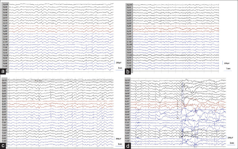
Figure 3.

(a-g): a) EEG showing left temporal focal spike appearance along with infrequent bilateral independent PSWC. b and c) PSWC appearing only during and post hyperventilation in graph “c” and absent before hyperventilation as noted in graph “b.” d) EEG showing decreased frequency of PSWC following sound stimuli and body movements
