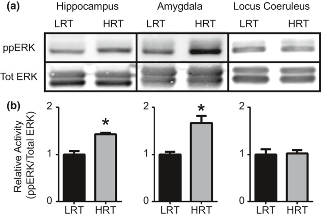
FIGURE 4.

Extracellular signal‐regulated kinase (ERK) activation. Western blot was performed to quantify ERK activation following 2 hr of physical restraint. Western blots for pERK and Total ERK (Tot ERK) from hippocampus, amygdala, and LC tissues (a). Quantification was performed using ImageJ software and statistically significant differences in pERK/TotERK levels are shown in (b) (*p‐value <0.05)
