
FIGURE 5.
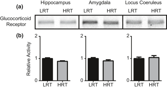
Glucocorticoid receptor. Western blot was performed to quantify GR levels following 2 hr of physical restraint. Western blots for GR from hippocampus, amygdala, and LC tissues (a). Quantification was performed using ImageJ software. No statistically significant differences were found for GR between LRT and HRT rats (b)