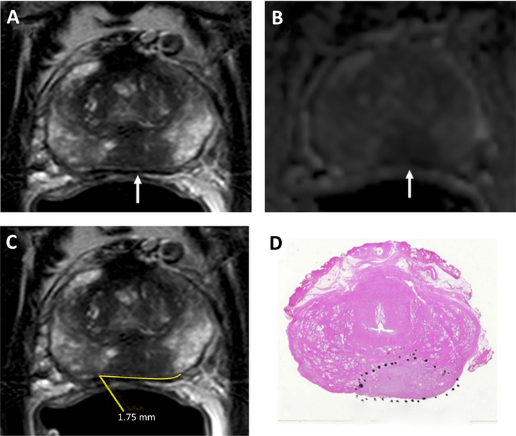
Fig. 1.
A 53-year-old man with a serum PSA = 23.70 ng/ml. Axial T2W MRI (A) and ADC map of DW MRI (B) show a midline to the left peripheral zone lesion (arrows), which represents a Gleason 3 + 4 prostate cancer lesion detected on TRUS-guided systematic prostate biopsy. The lesion does not have an overt extension but has a TCL of 17.5 mm (= 1.75 cm) measured on axial T2W MRI (C). Whole-mount prostatectomy specimen with H&E staining shows Gleason 4 + 4 prostate cancer within the lesion and posterior extraprostatic extension (inked in black) (D).

