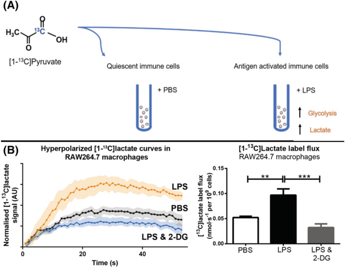
FIGURE 2.

A, in vitro activation of immune cells with pro‐inflammatory stimulus lipopolysaccharide (LPS) compared with quiescent immune cells given phosphate buffered saline (PBS). B, Hyperpolarized [1‐13C]pyruvate flux through LDH produces higher [1‐13C]lactate signals in LPS‐stimulated macrophages compared with control, unstimulated macrophages 53
