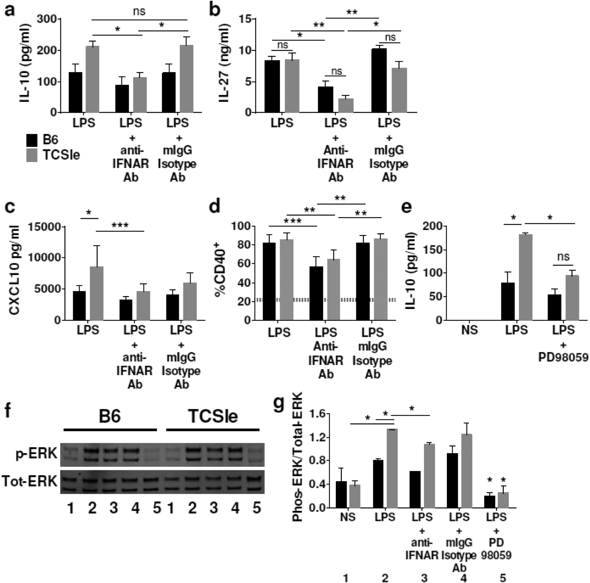
Figure 2.

Effects of IFNAR blockade on IL-10, IL-27 and ERK in TCSle cDCs in response to LPS. cDCs were treated with anti-IFNAR antibody or isotype control antibody for (a, b) 24 hours and then again for 30 minutes prior to stimulation with LPS or (c, d) one hour prior to stimulation with LPS. (a-b) Supernatants were collected 24 hours after LPS stimulation and (a) IL-10, (b) IL-27 and (c) CXCL10 were measured by ELISA. (d) cDCs were collected at 24 hours after stimulation and stained. Samples were gated on singlets, scatter gate, live cells, CD11c+ CD11b+, CD40+. Dotted lines represent CD40 expression in PBS treated B6 (20.6%) or TCSle (22.7%) (e) cDCs were treated with ERK inhibitor PD98059 for 30 minutes prior to LPS stimulation. Supernatants were collected 24 hours after LPS stimulation and IL-10 was measured by ELISA. (f) Representative Western blot and (g) averages and SD of the densitometries of Western Blots for phosphorylated ERK and total ERK. cDCs were treated with anti-IFNAR antibodies, or with ERK inhibitor PD98059 and LPS stimulation as above. Cells were harvested in ice cold western lysis buffer 1.5 hours after LPS stimulation. Western blots were probed for total and phosphorylated ERK. Number code: 1) Not stimulated, 2) LPS, 3) LPS + anti-IFNAR, 4) LPS + isotype control, 5) LPS + PD98059. (a-g) Averages and SD of 4–6 replicates from 3–4 experiments. (a-g) Statistical significance was calculated by ANOVA and post-hoc Tukey’s multiple comparison test. *p < 0.05, **p < 0.01, *** p < 0.001 ns means not significant.
