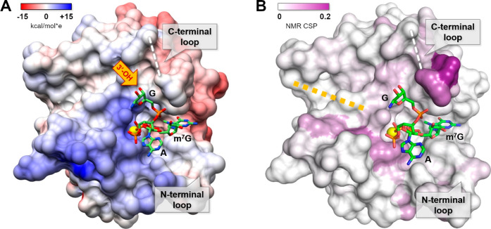
Figure 5.
Structure of trinucleotide cap analog [m7GppSpApG D2 (SP), chain A] in complex with eIF4E. The sulfur atom is represented as a yellow sphere, and the missing part of the C-terminal loop (the electron density was not well-defined in the X-ray structure) is marked by a white dashed line. The surface of the protein was colored according to Coulombic potential (panel A) calculated using the UCSF Chimera tool55 or color coded by 15N HSQC chemical shift differences between m7GpppApG and m7GTP bound eIF4E (panel B).

