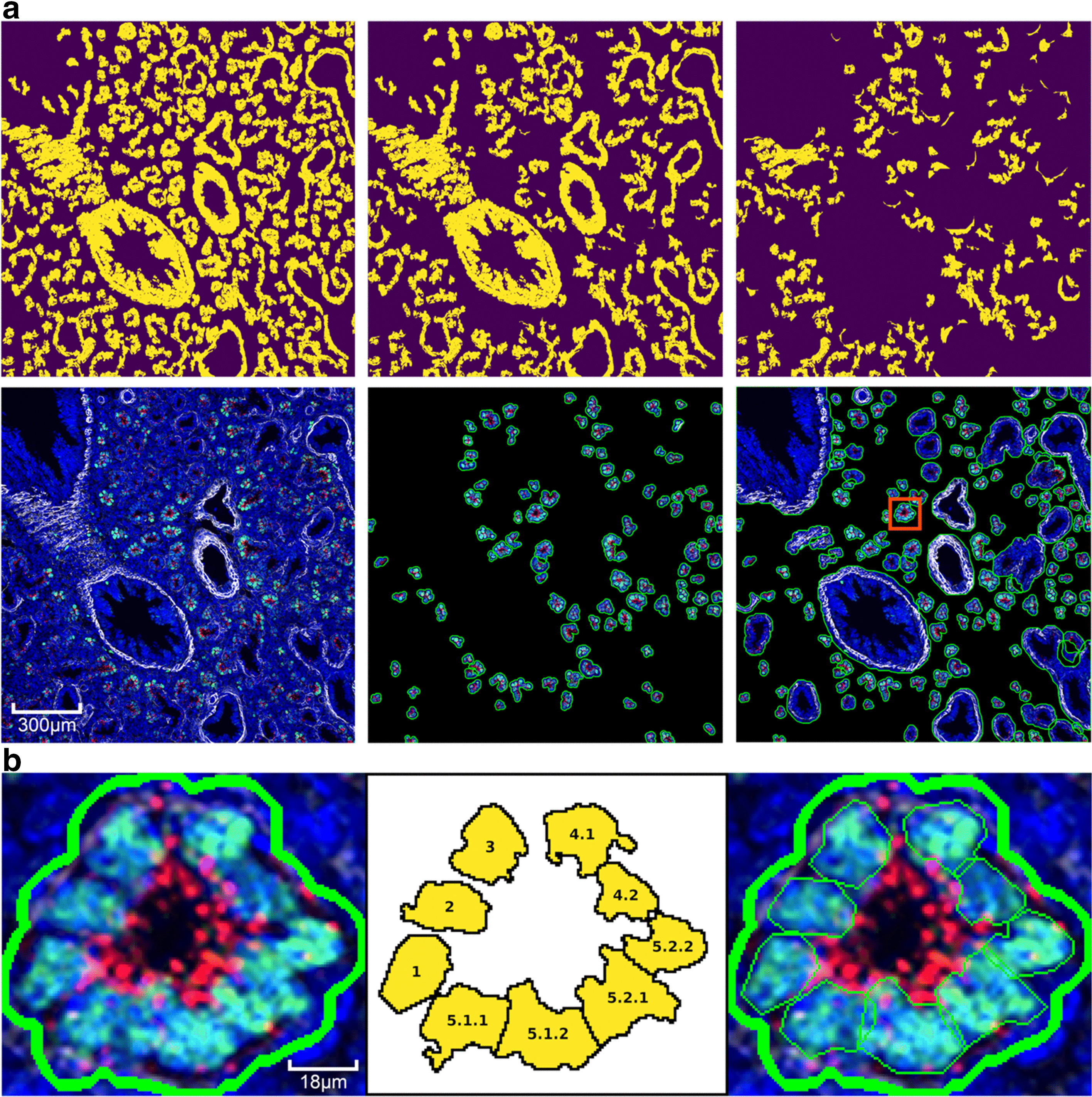
Fig. 6.

a Stages in automated structure segmentation of a mouse lung at developmental age E16.5. First row shows signal mask progression: top-middle after color segmentation stage, top-right is the residual signal mask after all stages. Second row: left shows original image, middle image shows candidate regions found in color segmentation stage, right shows all final candidates. The red box highlights a single region to demonstrate cell segmentation, b segmentation of cells within a distal acinar tubule with recursive spectral cluttering. From left to right: original structure from structure segmentation algorithm, order of segmentation of cells into structures in 3 recursive stages, cell segments within structure segment
