Figure 3.
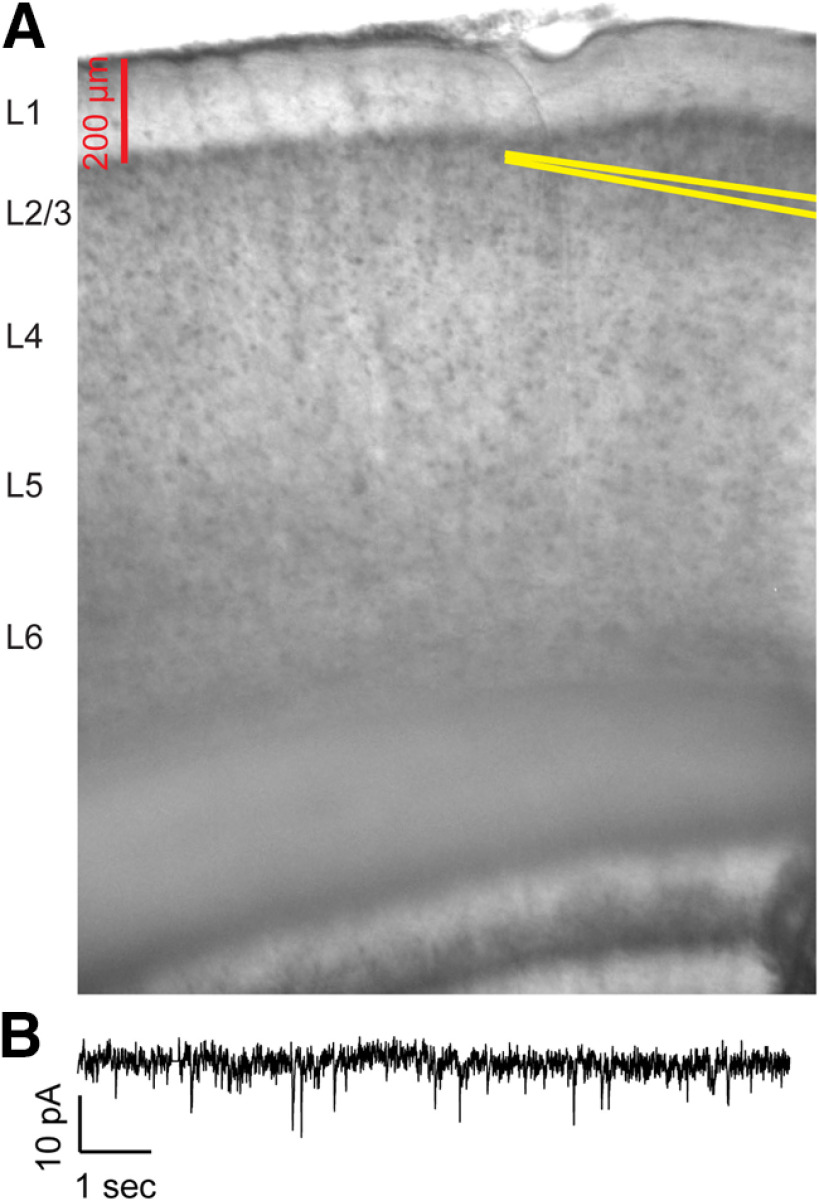
Image of auditory cortex slice and example voltage-clamp recording. A, Example image of recording electrode placement in a coronal slice containing auditory cortex. Recording pipette walls are highlighted in yellow lines. B, Example of membrane current recorded in voltage clamp with holding potential of −65 mV and bath application of 20 μm GABAzine.

