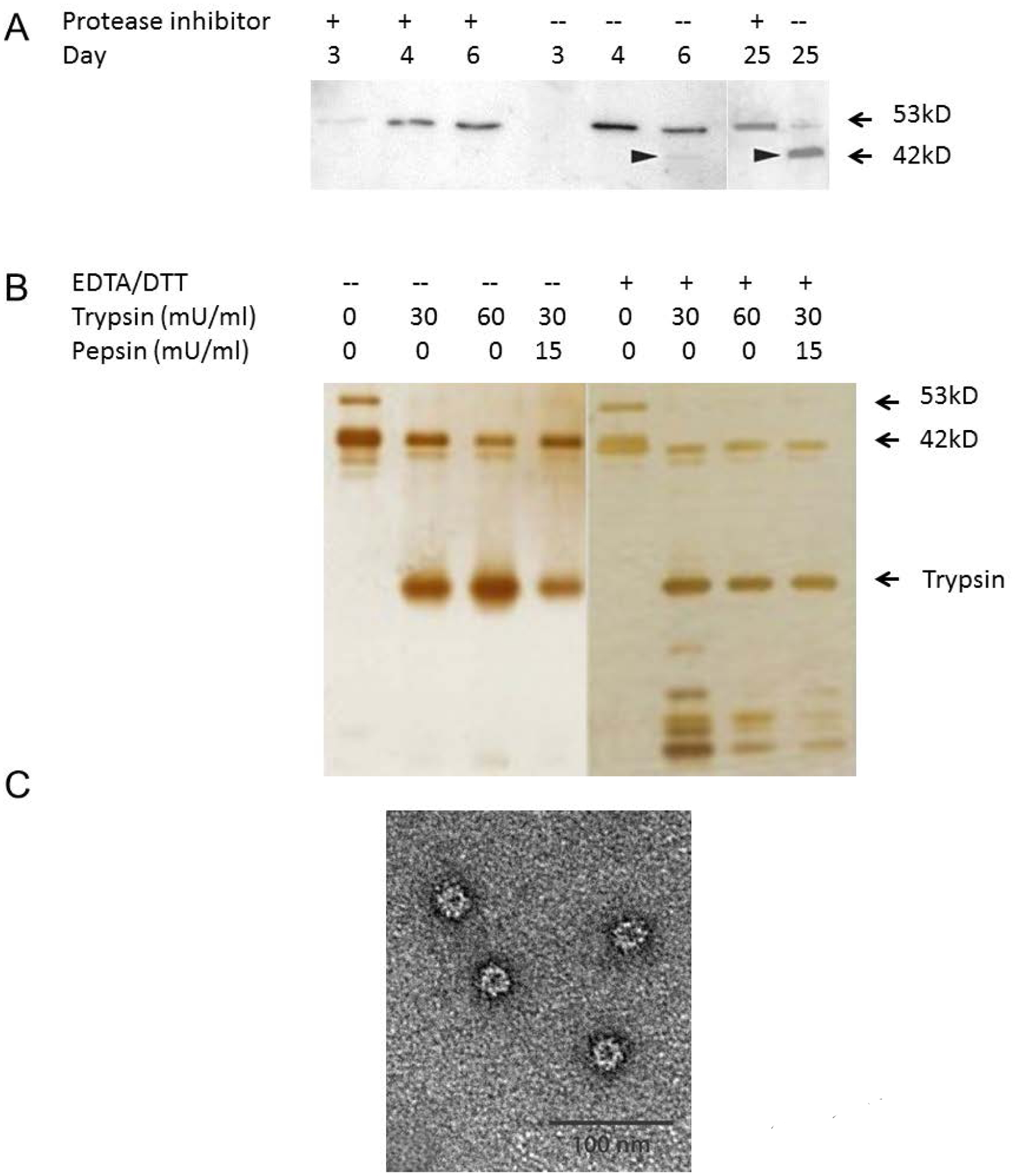
Figure 5.

Hydrolysis of the p18-VLPs. A: VLPs recovered from the culture media in the presence (+)/absence (−) of protease inhibitors were subjected on SDS-PAGE under reducing condition and then immunoblotted with anti-HIV antibody 447–52D. B: Eletrophoresis result of the p18-VLP pretreated with EDTA/DTT, 30mU/ml or 60mU/ml trypsin, and 15mU/ml pepsin, The SDS-PAGE was performed under reducing condition and developed with silver staining. C: Electron micrograph of negatively stained p18-VLPs after treatment with 60 mU trypsin. Bars = 100 nm.
