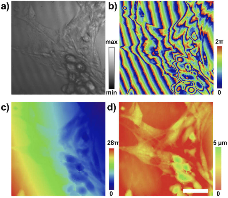
Fig. 5.
Phase sensitive OCM imaging of live fibroblast cell cultures. Imaging the surface of a collagen substrate to which the cells are attached permits sensing cumulative optical path differences induced by the cells. (a) The intensity component and (b) phase components are shown. Phase image is unwrapped (c) and flattened to produce quantitative images indicating the optical path delay induced by the cell (d). Representative images are shown with no averaging, captured at a 500 Hz framerate. Scale bar represents 20 .

