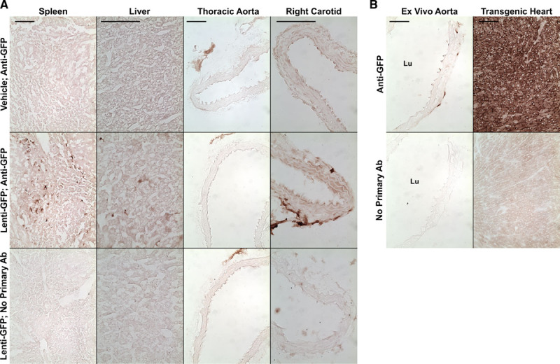
Figure 2.

Immunohistochemical detection of GFP (green fluorescent protein). Mice were injected with either vehicle or with a lentiviral vector expressing GFP. A, Sections of the indicated tissues were immunostained with an antibody (Ab) to GFP (Anti-GFP). Sections from mice injected with GFP-expressing lentivirus (Lenti-GFP) were also stained without the primary Ab. B, Positive control sections from an ex vivo–transduced aorta and from the heart of a GFP-transgenic mouse, with controls. Size bars=100 μm and apply to all panels in each column. Lu indicates lumen.
