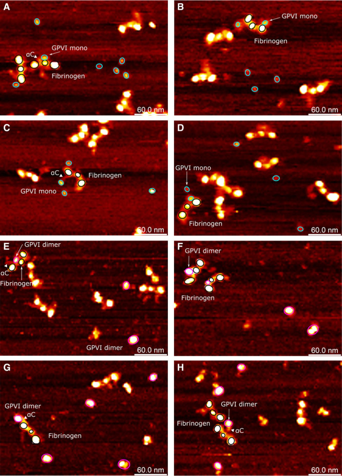
Figure 6.

Atomic force microscopy topography images of interactions between GPVI (glycoprotein VI) and fibrinogen. The fibrinogen molecules are highlighted using 3 black circles for its D-E-D structure. The disordered fibrinogen αC-regions are visible as flexible appendices with reduced height (and therefore reduced brightness) from the D-region or close to the E-region. A–D, GPVI monomer are indicated with cyan circles; (E–H) GPVI dimer are indicated with magenta circles. The αC-region of fibrinogen involved in GPVI binding is highlighted with triangles. Two structural isoforms of GPVI dimer were observed: one as a large single-domain globular protein and the other as an elongated shape or as 2 domains with similar sizes closely associated with each other.
