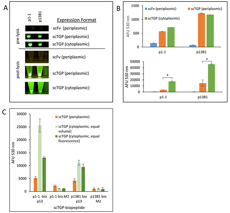
Fig. 3.

Assessing protein expression systems for scTGPs. The scFv format of the antibodies is expressed in the periplasmic protein expression vector, pEP. The scTGP (scFP) format of the constructs was expressed in both pEP (periplasmic expression) and pETCK3 (cytoplasmic expression). Panel A shows the fluorescence obtained after protein expression in both vector systems using the corresponding scFv constructs as controls. The upper panel B shows fluorescence from 100 μl of cell culture after two nights of protein expression (pre-lysis). The lower panel B shows fluorescence for equal volume of POP culture supernatant (post-lysis). The error bars were generated from three independent protein expressions. Panel C shows the functionality of scTGPs produced in the periplasmic and cytoplasmic compartments. Two scTGPs (p1-1 and p13B1) were analyzed by FLISA for binding to their cognate antigen (ITAM p13 peptide) using M2 peptide of influenza A as negative control. The activity of the scTGPs produced in the cytoplasm (green) was normalized to the activity of scTGP produced in the periplasm (orange) in two different ways: equal volume (light green) or equal fluorescence (dark green). * denotes a p value ≤ to 0.05. The error bars represent standard deviation generated from three independent binding assays.
