Fig. 2.
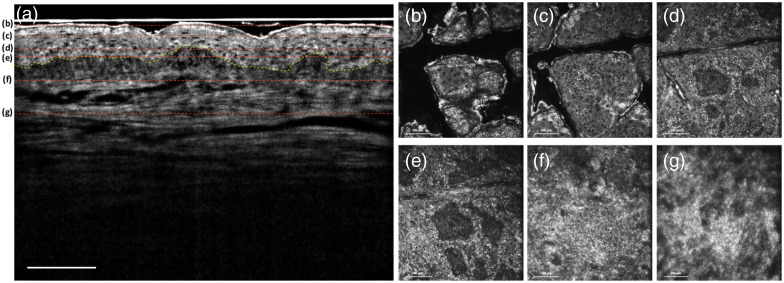
Normal skin: (a) in vivo FF-OCT B-scan image. The dashed yellow line presents the DEJ. (b)–(g) In vivo FF-OCT en face images at imaging depths of 30, 45, 65, 75, 110, and , respectively. The red dashed lines in (a) show the different depths of the en face images from (b)–(g). From (b)–(e), the different layers of epidermis including the stratum corneum, stratum granulosum, stratum spinosum, and stratum basale can be obtained. (f) and (g) The papillary dermis and reticular dermis, respectively, which have different structures of collagen fibers. Scale bar: .

