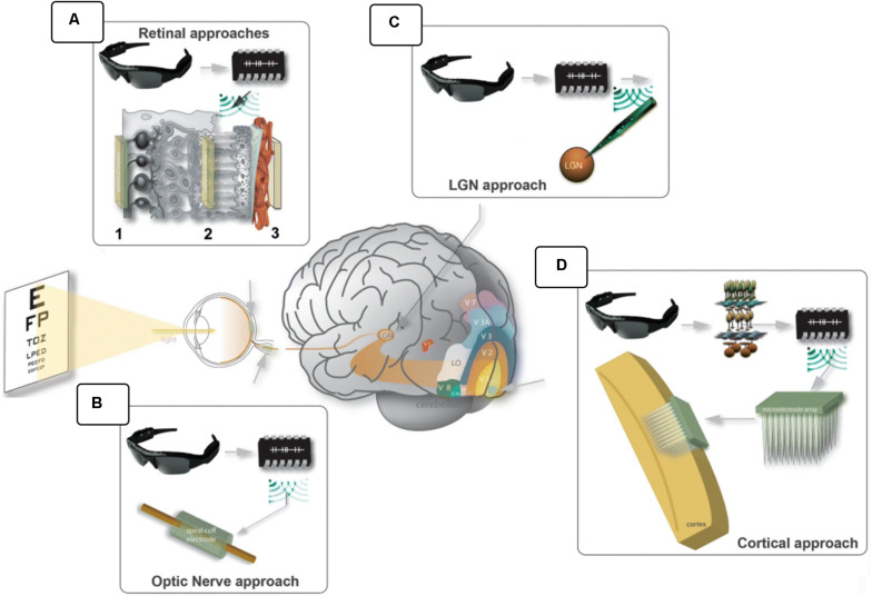
FIGURE 3.
Visual prostheses. In general, the scene is captured by a video camera, processed by a computer unit and sent to the electrical interface that stimulates the visual pathways. Different anatomical locations were explored: (A) epiretinal (1), subretinal (2) and suprachoroidal (3); (B) the optic nerve; (C) the lateral geniculate nucleus; and (D) the visual cortex. (modified from Fernandez, 2018; https://creativecommons.org/licenses/by/4.0/).

