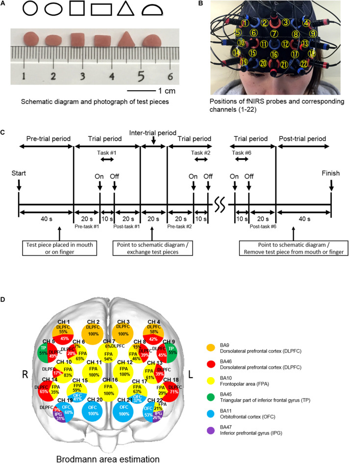
FIGURE 1.
Experimental aspects of finger and oral shape discrimination using prefrontal functional near-infrared spectroscopy (fNIRS). (A) Schematic diagrams and photographs of six types of test pieces: circle, ellipse, square, rectangle, triangle, and semicircle. (B) Positioning of the prefrontal fNIRS probes and corresponding channel numbers. (C) Timeline of a single session composed of six trials of shape discrimination in 8 min. (D) Location of the prefrontal fNIRS channels. Orange and red indicate the dorsolateral prefrontal cortex (DLPFC); yellow, the frontopolar area (FPA); green, triangular part of the inferior frontal gyrus (TP); blue, the orbitofrontal cortex (OFC); and purple, the inferior prefrontal gyrus (IFG). Each circle corresponds to a channel number. Pie chart shows percentage in cortical areas.

