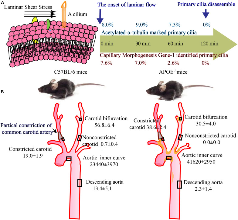
FIGURE 2.
Number of cilia in vitro and in vivo study. (A) The incidence of primary cilia is about 8% in static cultured ECs, gradually decreased with laminar flow and drop to 0% upon 2 h flow stimulation (Iomini et al., 2004). (B) Quantification of primary cilia per 0.005 mm3 in carotid and 0.5 mm3 in aorta. In WT and ApoE–/– mice, aorta and carotids were serially sectioned at 5 μm and then mounted to construct 3D image. Acetylated α tubulin was used to stain primary cilia and numbers of cilia per 0.005 mm3 in carotid and 0.5 mm3 in aorta are counted. Primary cilia are accumulated aortic arch, bifurcations, and constricted carotids, however, can nearly be detected in non-constricted carotids and descending aorta (van der Heiden et al., 2008).

