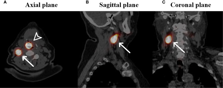
Figure 2.
Fused SPECT-CT images of a patient with a cT1N0 squamous cell carcinoma located on the laryngeal side of the epiglottis who underwent SN identification (own unpublished data). Arrowhead: primary injection site of 99mTc radioisotope, white arrows: identified SN in level II on the right side in the axial plane (A), sagittal plane (B) and coronal plane (C).

