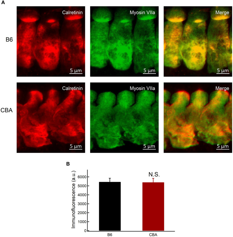
Figure 6.
Expression of calretinin in IHCs. (A) Whole-mount preparation of organ of Corti double-stained for calretinin and myosin VIIa. (B) Quantification of calretinin fluorescence at the base of IHCs in apical region without noise exposure. Statistical significance was assessed by unpaired t-test. N.S., no significant difference.

