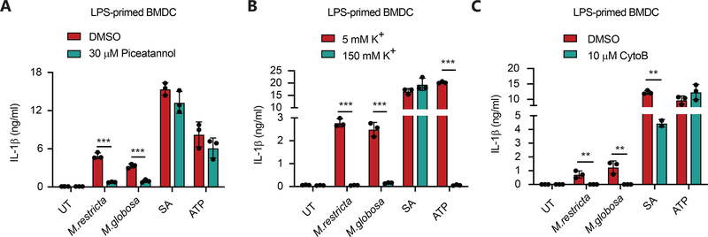
Figure 7. Malassezia-induced inflammasome activation is partially dependent on SYK signaling, potassium efflux and actin rearrangement.
(A) BMDC from wild type mice were primed with LPS (100 ng/ml, 4 hr) and stimulated with the indicated yeasts (MOI=5) for 3 hr or S. aureus (MOI=5) for 6 hr, or ATP (5 mM) for 30 min in the presence or absence of the SYK inhibitor Piceatannol. IL-1β secretion in the supernatants was measured by ELISA. (B) BMDC from wild type mice were primed with LPS (100 ng/ml, 4 hr) and stimulated as in (A) in the presence of the indicated levels of extracellular potassium. IL-1β secretion in the supernatants was measured by ELISA. (C) BMDC from wild type mice were primed with LPS (100 ng/ml, 4 hr) and stimulated as in (A) in the presence or absence of the actin polymerization inhibitor cytochalasin B. IL-1β secretion in the supernatants was measured by ELISA. All data represent measurements done in triplicate ± SD and statistics were done by student t-test * p≤0.05, ** p≤0.01,*** p≤0.001.

