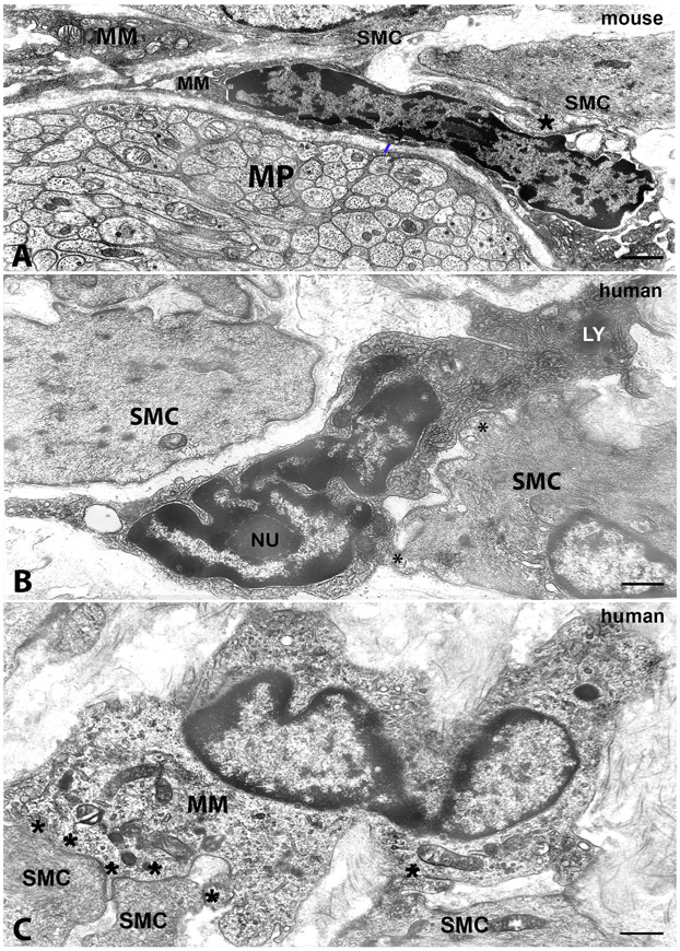
Figure 4. Transmission electron microscopic image. Morphology of macrophages and their cell-to cell contacts with smooth muscle cells.
A, mouse colon, myenteric plexus region. B, human stomach and C, human colon, circular muscle layer. A: MM: macrophage near a myenteric plexus ganglion (MP) and in close contact with smooth muscle cells (SMC). The asterisk indicates a MM-SMC contact of a peg-and socket type. The blue bar indicates a gap of 200 nm between the macrophage and the ganglion. MM has a dark cytoplasm and dark clumps of heterochromatin are accumulated along the nuclear envelope and close to the nucleolus. B and C: MM: macrophages in cell-to cell contacts (asterisks) with smooth muscle cells (SMC). In B, the MM has clumps of dark granular material, heterochromatin, adhering to the nuclear envelope all along the nucleus contour and around the nucleolus (Nu), a dark cytoplasm rich in coated vesicles and with several primary lysosomes and two dense bodies (LY). In C, the MM has a clear cytoplasm with several primary lysosomes and an indented nucleus. Scale bar: A,B: 0.6μm; C: 0.8μm.

