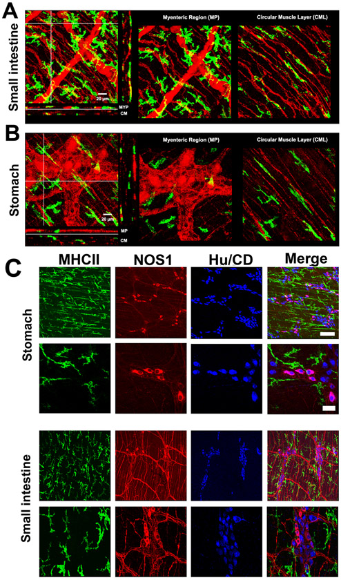
Figure 5. Muscularis macrophages and myenteric neurons in mouse.
A-B) 3D volume rendered images of MHC-II+ immunolabeling (green, macrophages) and PGP9.5 immunolabeling (red, neuronal structures) in small intestine and stomach from a 14-16 week old mouse. Scale bar: 20μm. Note that MHC-II+ cells are closely associated with PGP9.5 positive neurons in the myenteric ganglia and with nerve fibres at the sub-mucosal surface. In the mouse gastric body (B), MHC-II+ cells are also closely associated with PGP9.5 positive neurons in the myenteric ganglia but also observed within the circular muscle layer. C) Distribution of MHC-II+ macrophages in the stomach and small intestine in relation to NOS1 and HuC/D positive cells: low magnification, scale bar: 50 μm; high magnification, scale bar: 15 μm.

