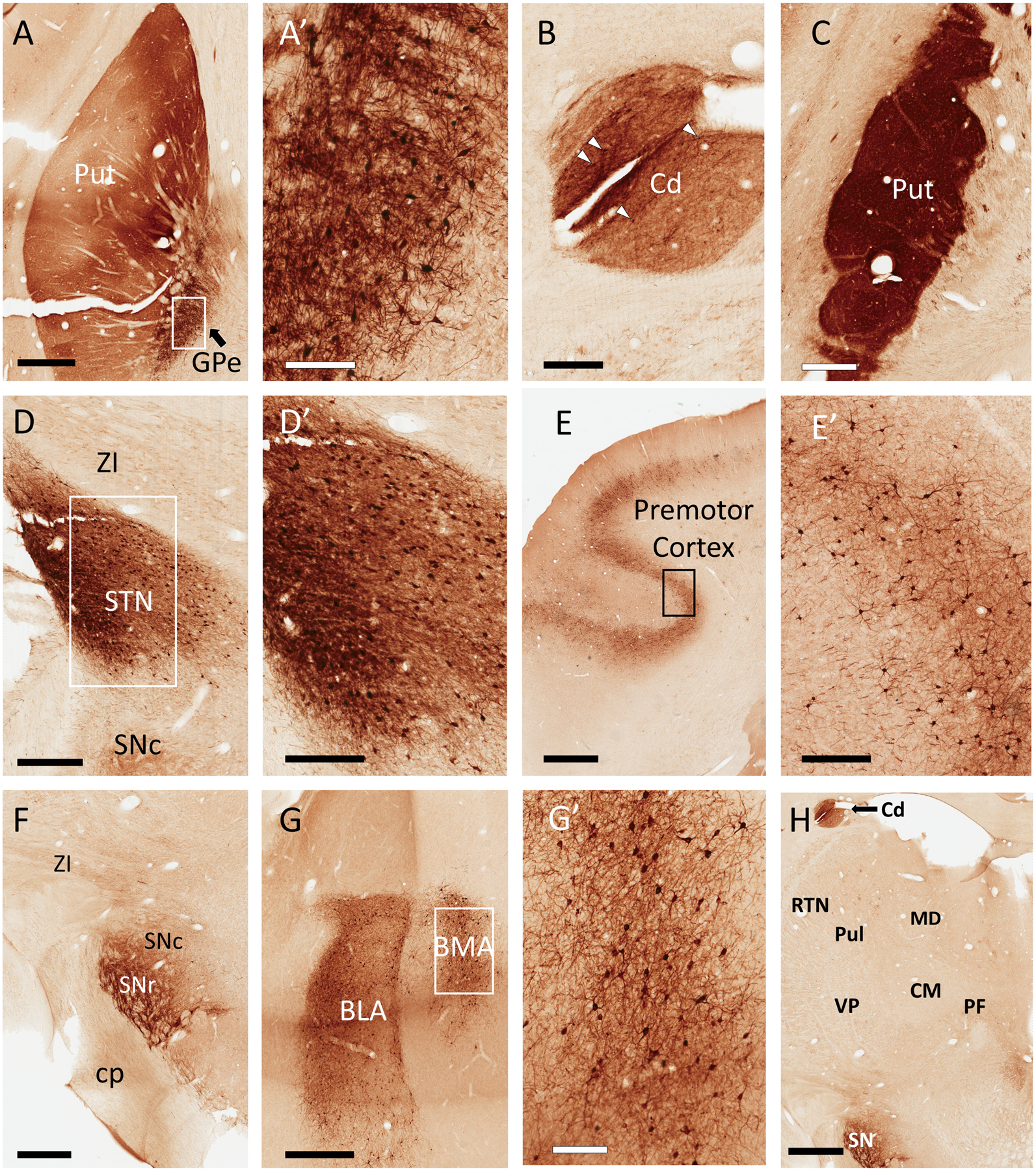
Figure 2:

Representative light micrographs of GFP expression in monkey brain following a unilateral, intra-striatal (putamen) injection of rAAV2-retro-hSyn-Jaws-GFP. All regions described below were ipsilateral to injection site. In the putamen (Put), neuropil was diffusely labeled, with scattered GFP+ cell bodies, particularly in the ventral territory (A). In the same section, numerous GFP+ cell bodies were found in the external globus pallidus (GPe) (A and A’). Similar to the Put, GFP+ fiber staining was observed in the caudate (Cd) (B; note that this section of caudate is also visible in Panel H). Similar to the rat striatum, the neuropil of the Put was densely labeled across the rostrocaudal axis, many millimeters away from the injection site (C). The subthalamic nucleus (STN) contained numerous GFP+ cell bodies and dense neuropil staining (D and D’). Varying numbers of deep-layer pyramidal cells were labeled in many cortical regions, including the premotor cortex (E, E’). In the substantia nigra, we observed GFP+ fibers, as well as a few labeled cell bodies bordering the compacta (SNc) and reticulata (SNr) territories (F). Numerous GFP+ cells were located in the basolateral and basomedial amygdala (BLA and BMA, respectively) (G and G’). Remarkably, the thalamus was nearly devoid of GFP labeling (H). Additional abbreviations: centromedian nucleus of thalamus (CM), parafascicular nucleus of thalamus (PF), pulvinar (Pul), thalamic reticular nucleus (RTN), ventral posterior nucleus of thalamus (VP), zona incerta (ZI), all other abbreviations as described in Figure 1. Scale Bars: 2mm (E, H) 1mm (A, F, G); 600μm (C); 500μm (D); 300μm (B, D’, E’); 200μm (A’, G’). Approximate rostrocaudal coordinates of sections, according to (Paxinos et al., 1999) in mm relative to interaural line: 10.2 (A, D); 7.5 (B, C, F, H); 19.65 (E); 14.7 (G). Figure includes micrographs from both monkeys used in this study.
