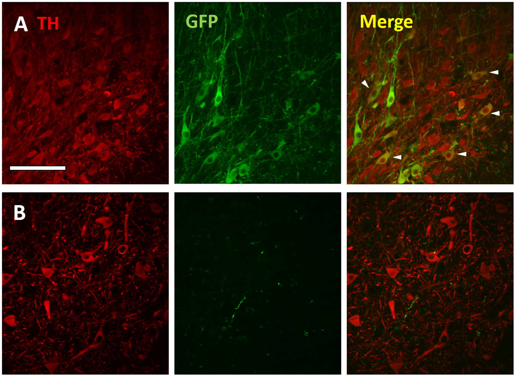
Figure 3:

GFP expression in dopaminergic (tyrosine hydroxylase (TH)-positive) neurons in the rat, but not monkey SNc following intra-striatal rAAV2-retro injection. Confocal images of TH (red; left), GFP (green; middle) and merged markers (right) of the rat (row A) and monkey (row B). SNc ipsilateral to injection site. Arrowheads in merged image depict a subset of GFP+/TH+ cells (yellow) in the rat SNc. Scale bar: 100μm.
