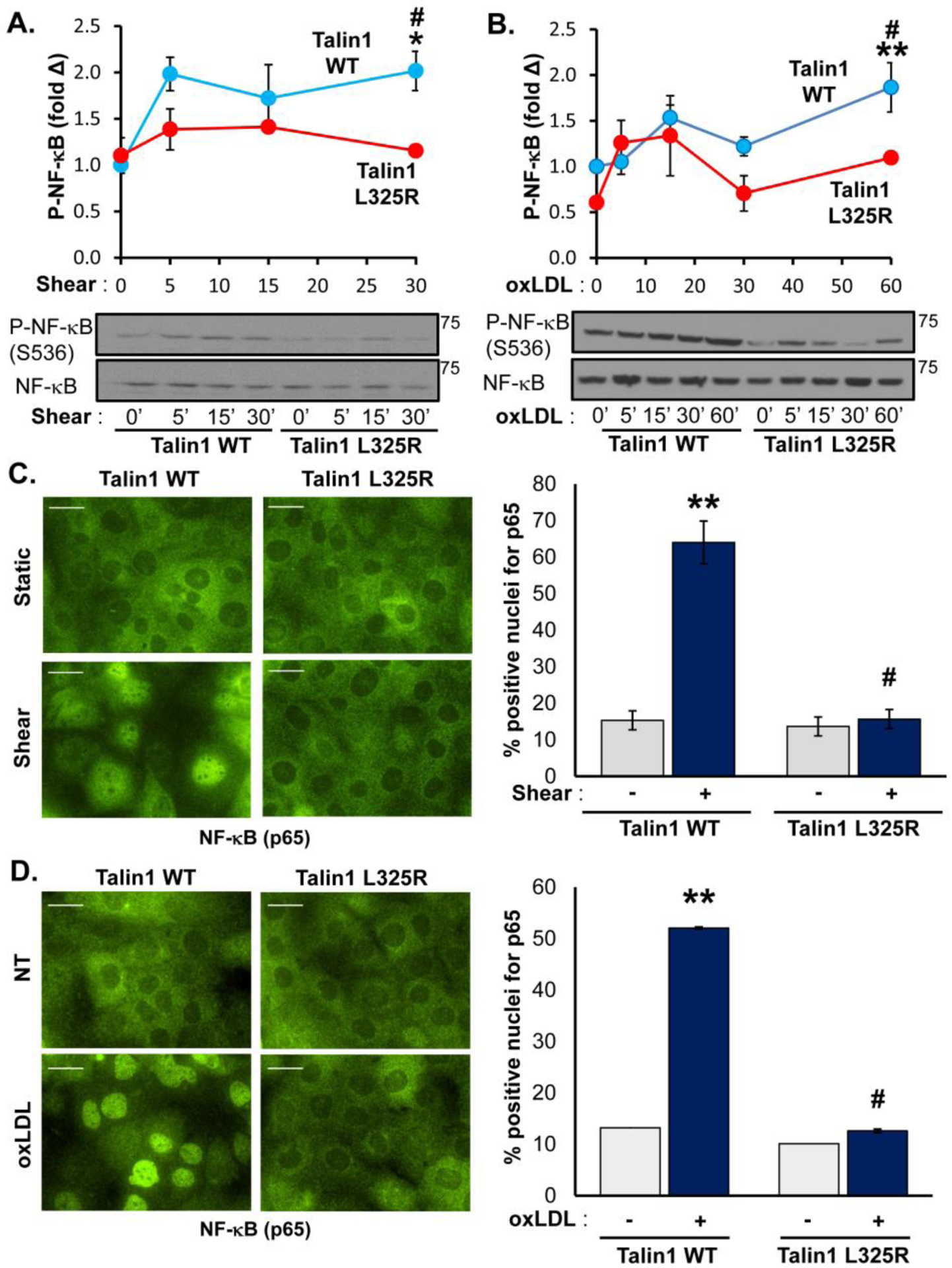
Figure 3. Blocking Talin1-Dependent Integrin Activation Blunts Shear- and OxLDL-Induced NF-κB Activation.

A/B) Talin1 WT and talin1 L325R MLECs were plated on fibronectin and either (A) exposed to onset of laminar flow (shear) or (B) treated with oxLDL (100 μg/ml) for the indicated time points. NF-κB activation was assessed by Western blotting for NF-κB phosphorylation (P-NF-κB (p65, Ser536)). Representative Western blots are shown. n=4–5. C/D) Talin1 WT and talin1 L325R MLECs were treated with either (C) shear for 30 minutes or (D) oxLDL for an hour, and NF-κB activation was assessed by immunocytochemistry for NF-κB nuclear translocation. At least 100 cells were assessed per experiment. Representative images are shown. n=4. Scale bar = 25 μm. Values are means ±SE. *p<0.05, **p<0.001 indicates significance compared to untreated controls, and #p<0.05 indicates significance between talin1 WT and talin1 L325R MLECs. Statistical analysis was performed using 2-way ANOVA with Bonferroni posttest.
