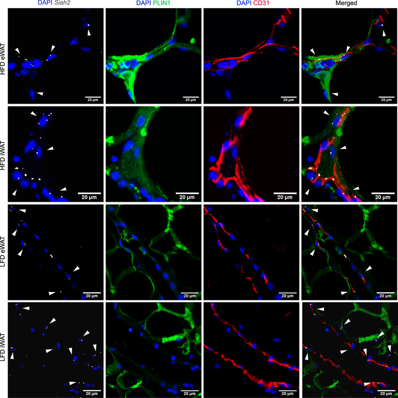
Figure 2:
Siah2 localizes near the adipose tissue vasculature. Representative images of paraffin embedded inguinal (iWAT) and epididymal (eWAT) adipose tissue sections from male mice fed a 60% high fat diet (HFD) or 10% low fat diet (LFD) were stained for nuclei (DAPI, blue), Siah2 mRNA (gray), and PLIN1 (green). CD31 (red), a marker of endothelial cells, was used to visualize the vasculature endothelial cells. Signal brightness of everything except Siah2 was dimmed in the merged image to enhance Siah2 visualization. The arrows are added to emphasize Siah2 localization. Data represent observations from N=8/group.

