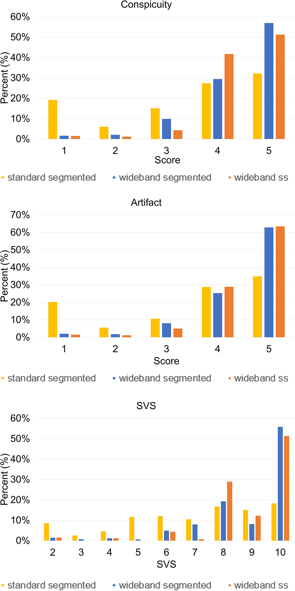
Figure 3:

Distribution of conspicuity, artifact and SVS for three LGE pulse sequences. Standard segmented LGE produced considerably more non-diagnostic images than both wideband LGEs. SVS: summed visual score.

Distribution of conspicuity, artifact and SVS for three LGE pulse sequences. Standard segmented LGE produced considerably more non-diagnostic images than both wideband LGEs. SVS: summed visual score.